Incomplete Abortion
- Products of conception are not completely expelled out
Clinical Features:
- History of per vaginal fleshy mass expulsion
- Continuation of abdominal pain
- Continuation of per vaginal bleeding
Ultrasound Findings:
- Irregular Mixed echogenic substances are noted within the uterine cavity
Case 01:
Case 02:
Case 03:
Case 04:
Case 05:
- Vascularity may be noted on Doppler if trophoblast tissue is present. Urine for PT may become positive there
Case 06:
- In early gestation, irregular distorted or displaced gestational sac along with pervaginal bleeding may indicate incomplete abortion
- Proper views for GSD or MSD is not be possible at this situation
Case 07:
Case 08:
Hydrops Fetalis
Ultrasound Findings:
- Fluid accumulation in 2 or more body cavites.
- May be immune or non-immune
Case 01:
Fetal Unilateral Hydronephrosis
Ultrasound Findings:
- Fetal unilateral enlarged kidney with gross hydronephrosis without any hydroureter.
- Urinary bladder appears normal and dynamic.
Case 01:
Polycystic Ovarian Disease (PCOD)
Clinical Features:
- Subfertility
- Irregular scanty menstruation or amenorrhoea
- Obesity
- Hirsutism
Ultrasound Findings:
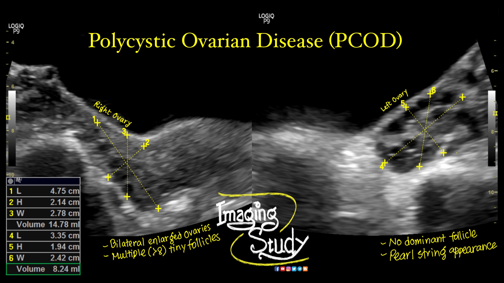
- Bilateral enlarged ovaries
- Multiple tiny cystic follicles (>8) at peripheral parts of both ovaries forming 'Pearl-String appearance'
- Echogenic stroma
- No dominant follicle
- Thickened endometrium may be seen
Case 01:
Cord Around The Fetal Neck
- Check the number of umbilical vein for better understanding the layers
- Always use color Doppler to assess the neck in late gestation.
- 3 layers of cord around the neck
Case 02:
- 3 layers of cord around the neck
Colloid Nodule of Thyroid Gland
- Cystic nodule with in the thyroid parenchyma.
- Echogenic foci casting comet tail artifacts are seen with in the nodule.
Testicular Torsion
- Swollen testis & epididymis.
- Homogeneous or heterogeneously hypoechogenic parenchyma.
- Intratesticular anechoic area of necrosis.
- No intratesticular venous flow.
- Absent or reduced intratesticular arterial flow.
- Peritesticular increased arterial flow.
- Mild reactionary hydrocele.
- Thickened scrotal skin.
Cholecystectomy Clips Ultrasound
- Bright straight echogenic structure at gall bladder fossa.
- May casts acoustiic shadow.
- History of laparoscopic cholecystectomy.
Pleural Effusion Ultrasound
- Abnormal collection in pleural cavity.
- May be clear or hazy, with or without septaion depending on type.
- Loss of mirror image artifact.
Mild Fatty Change in Liver (Grade I)
- Increased hepatic parenchymal echogenicity.
- Increased attenuation of ultrasound beam.
- Well visualized intrahepatic vessels.
- Well visualized diaphragm.
- May or may not be associated with hepatomegaly.
- Common in obesity, diabetes, pregnancy.
Adenomyomatosis of Gall Bladder
- Focal gall bladder wall thickening.
- Comet tail artifact.
- Twinkling artifact on color Doppler.
Gall Bladder Polyp
- Hyperechogenic structure/s attached to the gall bladder wall.
- No acoustic shadow.
- Thickness upto 10 mm.
- Not moves along posture changing/ Fixed
- Peduncle is well visualized if magnified
- Commonly missed if not searched using 'Zoom' option
Cholelithiasis
- Luminal hyperechogenic structure/s
- Casting posterior acoustic shadow/s
- Moving or not moving along the posture changing
- May be associated with cholecystitis
- Common in Female, Fatty, Fertile, Forty, Fair (5F)
Elongated Gall Bladder
- Length is more than 10 cm.
- Normal anatomical variety.
- Asymptomatic patient.
- Sometimes it may extend up to the right iliac fossa.
Hepatic Metastasis
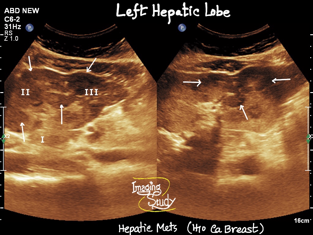
- Hypo or hyperchogenic space occupying lesions
- Ill defined
- Internal vascularity on color Doppler
- May compress adjacent vasculature
- Search for the primary source